, its stages and meaning.")


 review: Gorgeous display meets Ryzen power")


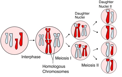

What is meiosis?
- The term meiosis is derived from the Greek word ‘half which means reduce.
- It is a special type of cell division that occurs in organisms that reproduce sexually. It occurs only in the gonads (testes and ovaries in animals and anthers and ovules in higher plants) to produce gametes.
- It also occurs in sporangial sacs to produce sporangiospores in lower plants and fungi that reproduce asexually by sporulation.
- Meiosis is a type of cell division in which a mother cell divides to produce four haploid daughter cells with half the normal number of chromosomes than in the mother or progenitor cell itself..
- Since the number of chromosomes in the daughter cells is reduced by half, the process is also called reductional cell division. For example, a diploid human cell has 46 (23 pairs) chromosomes, one set (23) from the male gamete and another set (23) of chromosomes from the female gamete.
- When opposite gametes fuse, a diploid zygote is formed that divides by mitosis. So, each cell in the human body has 46 chromosomes, except gametes. However, men’s sperm and women’s eggs only have 23 chromosomes.
- In each diploid cell, there are two identical chromosomes that form a pair, called a homologous chromosome pair. There are 23 pairs of homologous chromosomes in a diploid human cell. Only one chromosome of each pair is present in a haploid cell (gamete).
How many nuclear divisions occur in meiosis?
- Meiosis consists of two successive nuclear divisions with a single doubling of the amount of DNA or chromosomes. These divisions are called the first and second meiotic divisions.
- During the first meiotic division, homologous chromosomes pair. The pair then separate and one member goes to each of the daughter cells. It is important to note that the centromere does not divide. This division reduces the number of chromosomes in the mother cell by half. The two daughter cells thus formed have half the chromosomes contained in the mother cell. Therefore, this division is also called reductional division.
- During homologous pairing, each chromosome within a pair can exchange DNA. The exchange of genetic material between non-sister chromatids of a homologous pair occurs at specific contact points called chiasmata (plural: chiasm) and is known as crossing. Due to crossing over, the newly formed chromosomes are unique and create genetic variation in the gametes produced. Each of the four cells produced by meiosis is genetically different from each other and from the mother cell.
- In the second meiotic division, the chromatids of each chromosome separate at the centromere and move to opposite poles. The separated chromatids are now called new chromosomes. When chromosomes reach opposite poles of the cell, new nuclear membranes form around them. Each daughter cell divides in two, forming four haploid cells in total.
- During spermatogenesis (sperm formation) In men, four sperm are produced from one stem cell during meiosis, while in women, during oogenesis (formation of an egg), four daughter cells are produced from one mother cell, however, only one of them becomes an egg and the other three degenerate.
Meiosis I (First meiotic division):
- Meiosis, like mitosis, also consists of five stages; Interphase, Prophase I, Metaphase I, Anaphase I and Telophase I.
- It is a heterotypic division (which reduces the number of chromosomes from diploid to haploid).
- Interface:
- Prepares the cell for successful division.
- As in mitosis, the cell increases in size.
- Copying of all chromosomes occurs and the nucleus has distinct membranes.
- Prophase I:
- The first meiotic prophase is extremely long in duration compared to mitotic prophase and consists of the following five sequential substages; Leptotene, Zygotene, Pachytene, Diplotene and Diakinesis.
- leptotene:
- The nucleus is large and increases in size. The nucleolus is prominent.
- The chromosomes are coiled and therefore become visible as threadlike, intermingled structures.
- leptotene:

-
-
- Chromosomes are double-stranded even though they appear single-stranded.
- Chromosomes have a bead-like appearance due to the fact that chromatin is wound around histone proteins to form nucleosomes. The number, size and position of genes are constant and identical in homologous chromosomes.
- In animal cells, the centrosome divides and moves to opposite poles in the form of centrioles.
- zygotene:
- Homologous chromosomes (one maternal and one paternal) approach each other and form pairs called homologous pairs. This pairing of homologous chromosomes is called synapse. Each pair of homologous chromosomes at synapses is called bivalent.
-

-
-
- The pairing of homologous chromosomes is very precise and involves point-to-point pairing along the entire length of the homologous chromosomes.
- Chromosomes undergo more coiling and condensation. They become shorter and more clearly visible.
- The nucleolus increases even more in size and the centrioles become more separated.
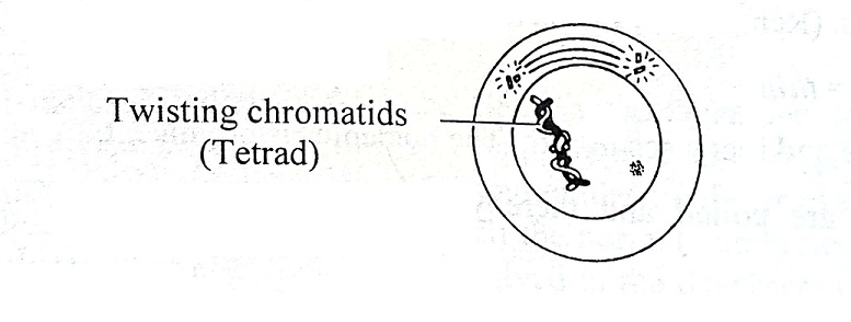
- pachytene:
- The paired chromosomes of each bivalent become shorter, thicker, and clearly distinct. At this stage, the synapse is completely completed.
- Each chromosome divides longitudinally (lengthwise) into two sister chromatids. Each pair of chromosomes now consists of four chromatids and is called tetrad.
- The non-sister chromatids of a tetrad twist around each other and touch each other at different points of contact.
-
-
-
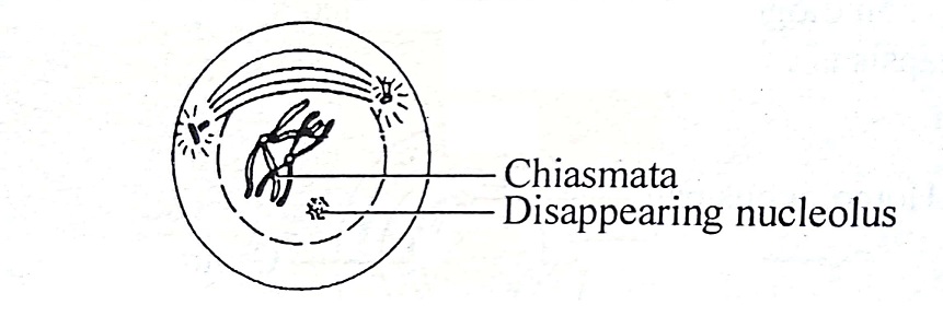
- An exchange of chromatid segments occurs between the two non-sister chromatids of each homologous pair after coiling around each other. this is called crossing. During crossing over, the non-sister chromatids of a tetrad break at identical points. The broken segment of a chromatid joins with the non-sister chromatid of its tetrad at different points. This point of exchange and reunification of the chromatids is ‘X’ shaped and is called chiasm (plural: chiasmata). Simply put, crossing over occurs at the chiasm.
- diplotene:
- The paired homologous chromosomes begin to separate from each other due to a repulsive force and begin to move away from each other.
-
-
-
- Non-sister chromatids that separate from a homologous pair remain joined at some points called chiasm.
- The nucleolus and nuclear membrane begin to disappear.
- Diakinesis:
- Further shortening and condensation of chromosomes occurs. Chromosomes move toward the periphery of the nucleus.
- Chiasmata moves towards the end of the chromosomes. This process is called termination of chiasmata.
-

-
-
- The nucleolus and nuclear membrane continue to disappear, marking the end of prophase I.
- In animal cells, centrioles reach their respective poles of the cell and begin to form spindles.
-
- Metaphase I:
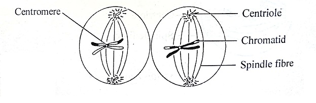
- The chromosomal tetrads (four chromatids) are located in the equatorial plane of the spindle fibers.
- Spindle formation is complete and several spindle fibers are attached to the centromeres of the chromosomes.
- The nuclear membrane and nucleolus disappear completely.

- Anaphase I:
- The two chromosomes of a homologous pair separate from each other and move toward opposite poles of the cell as they separate by the shortening of the spindle fibers. Unlike anaphase of mitosis, the centromeres of each chromosome do not divide.

-
- At the end of anaphase I, two groups of haploid chromosomes form, one at each pole of the spindle. The consequence of this stage of meiosis is the reduction of the number of chromosomes by half compared to diploid. However, the chromatids of these chromosomes are not genetically identical due to crossing over. This is the essential difference between meiosis and mitosis. The chromosomes at the poles are from the father or the mother.
- Telophase I:
- At each pole, the chromosomes lose their identity and become a network of chromatin.
- The nucleolus disappears completely.
- The nuclear membrane reappears around the chromatin network and two daughter nuclei are formed.

-
- Each daughter nucleus thus formed has half the number of chromosomes that the nucleus of the mother cell has. This condition is called haploid (n). Therefore, two haploid nuclei are formed, followed by cytokinesis that results in the formation of two haploid daughter cells.
Meiosis II (Second meiotic division):
- The interface between the first and second meiotic division is absent or if present it is of very short duration.
- The two haploid cells formed by the first meiotic cell go through four substages; Prophase II, Metaphase II, Anaphase II and Telophase II.
- This second meiotic division is a mitotic division They form daughter cells with the same number of chromosomes as the mother cell, so called. homotypic division.
- The changes occur simultaneously in both haploid cells.
See also: Differences between mitosis and meiosis
- Prophase II:
- The chromatin network changes to chromosomes due to condensation. The nuclear membrane and nucleolus disappear.
- Each chromosome consists of two distinct chromatids attached to a single centromere.
- The centriole duplicates and moves to opposite poles.
- Spindle fibers begin to appear.

- Metaphase II:
- Spindle formation is complete. Chromosomes, each of which has chromatids, are arranged along the equatorial plane of the spindles.
- The centromere of each chromosome attaches to the spindle fibers.
- Anaphase II:
- The centromere of each chromosome divides and separates so that each chromatid (which now behaves like a chromosome) has its own centromere.
- The separated chromatids move away from each other towards opposite poles which are now called chromosomes.

- Telophase II:
- The chromosomes in each group become long and thin and form a network of chromatin.
- The nuclear membrane is formed around them and they are organized into a nucleus.
- The spindle fibers disappear and the nucleolus reappears.

Cytokinesis:
- Nuclear division is followed by cytokinesis. It begins in Anaphase II and ends in Telophase II.
- The centriole duplicates to form a centrosome in each daughter cell.
- Cytokinesis is completed with the formation of four daughter cells. Each cell has a haploid number (n) of chromosomes.
- Therefore, meiosis results in the formation of four haploid daughter cells from a single diploid (2n) mother cell.
Importance of meiosis:
- Stability of living beings (species):
- Reduces the number of chromosomes by half in daughter cells. Gametes are formed as a result of meiosis. Gametes fuse during fertilization and restore the number of diploid chromosomes. Thus, it maintains the number of chromosomes from generation to generation in organisms that reproduce sexually. In other words, it prevents the multiplication of the number of chromosomes and maintains the stability of the race of the species.
- Genetic difference (Variation):
- Through crossing over, non-identical cells are formed, that is, gametes. This brings variation in organisms of the same species. This variation results in long-term evolution.
Meiotic cell division (Meiosis), its stages and meaning.
#Meiotic #cell #division #Meiosis #stages #meaning