")

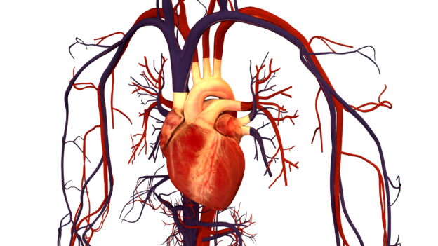

- He blood vessels that transport oxygenated blood away from the heart and supply blood to various parts of the body constitute the arterial system.
- The main blood vessels that arise from the left ventricle are called systemic aorta.
- This aorta is branched to form many smaller arteries, which in turn branch many times in arterioles.
- The aorta runs up and curves abruptly or arches down. The aorta ascending career portion is called ascending aorta, And the aorta down is called descending aorta.
1. Ascending aorta: Enter:
- Coronary arteries: There are two coronary arteries (right and left) that arise from the base of the aorta. They supply oxygenated blood to the walls of the heart.
2. Aortic arc emits the following arteries:
to. Innominated artery or brachiocephalic trunk:
b. Left Common Carotid Artery:
do. LEFT SUBCLAVIA ARTERIA:
- INNOMED ARTERIA (Brachiocephalic): It has approximately 4 to 5 cm long, which is soon divided into two branches to form the common carotid artery and the right subclavian artery.
- Right common carotid artery: Run up on the side of the trachea and enter the neck and head. It is divided into external and internal carotids that supply blood to the right side of the head and face.
- Right subclavian artery: Emits the following arteries:
- Vertebral artery: It supplies the head and parts of the right shoulder.
- Right axillary artery: Supplies blood to the scapular region.
- Right brachial artery: Supplies blood to the arm.
- Right radial artery: It supplies the front arm, the elbow joint, the radio and the muscles on the side of the forearm assault.
- Right cover artery: Supplies the cubito, the forearm, the muscles of the ulnar side of the forearm.
- Left Common Carotid Artery: It arises directly from the aorta. Run up on the side of the neck (trachea). It is divided into external and internal carotid arteries.
- He internal carotid artery supplies blood to most of the brain. It also supplies blood to the eyes, forehead and nose.
- He external carotid artery They branch more to supply blood to various parts of the body as follows:
- Upper thyroid artery: Supplies blood to the thyroid gland and adjacent muscles.
- Lingual artery: Supplies the tongue, mouth floor, etc.
- Facial artery: Supplies the facial parts of the mouth.
- Occipital artery: Supplies the back of the head.
- Temporary artery: It supplies the front, temporal and parietal parts of the scalp.
- Maxillary artery: Supply the muscles of the mouth.
- LEFT SUBCLAVIA ARTERIA: It arises directly from the aorta and is divided in the same way that the right subclavian branch of the unnamed artery. It supplies blood to the body parts similar to which the right subclavian artery is performed but to the left side.
- Vertebral artery: Supplies the neck and parts of the left shoulder.
- Left axillary artery: Supplies the scapular region.
- Left brachial artery: It supplies the upper forearm, the humerus and the elbow joint. The brachial artery in the previous elbow is used to measure blood pressure.
- Left radial artery: It supplies the front arm, the elbow joint, the radio and the muscles on the side of the forearm assault.
- LEFT COBITAL ARTERIA: Supplies the cubito, the forearm, the muscles of the ulnar side of the forearm.

3. Thoracic aorta:
The chest segment of the descending aorta (also called thoracic aorta) emits several pairs of small arteries and supplies blood to the following parts:
to. Bronchial arteries (not paired): They supply the bronchi, bronchioles and connective tissues in the lungs.
b. Esophagial arteries (not paired): They supply the esophagus.
do. Intercostal arteries:
- Dorsal branch (9 pairs): They supply the spinal cord, muscles and back skin.
- Subsequent intercostal arteries (1 torne under rib 12):
- Muscle branch: Supply the chest muscles.
- Cutaneous branch: Supplies the thorax skin.
- Breast branch: Supplies the breast glands of the breasts.
d. Superior phrenic arteries: They supply to the diaphragm.
4. Abdominal aorta:
After drilling the diaphragm, the descending arch of the aorta becomes abdominal aorta and emits a series of matched arteries and not paired to supply different parts of the body as follows:
to. Lower phrenic arteries: They supply to the diaphragm and the lower esophagus.
b. Celiac artery (trunk): It is not separated and originates immediately below the diaphragm and emits three branches:
- Left gastric artery: Supplies blood to the stomach.
- Common Hepatic Artery: It supplies blood to the liver, the gallbladder, the pancreas and the pyloric stomach.
- Splenic artery: Supplies blood to the parts of the pancreas, stomach, omentum and spleen.
do. Upper mesenteric artery (not paired): It is a coarse and long non -paired artery that is further branched as:
- Lower pancreaticodenal artery: It supplies blood at the head of pancreas and duodenum.
- Yeyunales and ileal arteries: SUPPLY TO THE DELGADO INTESTIN.
- Ileocolic artery: Supplies to the lower ileum, the appendix, the ascendant and part of the cross colon.
- Right colic and medium colic arteries: Supply to the transverse and ascending colon.
d. Lower mesenteric artery (not paired): It branches even more like:
- Left colic artery: Supplies to the transverse and descending colon.
- Sigmoid artery: Supplies to descend and sigmoid colon.
- Upper rectal artery: Supplies blood to the rectum and the anal region.
my. Renal arteries: A pair of renal arteries supply blood to a couple of kidneys. Renal arteries also emit adrenal arteries that supply blood to the adrenal (adrenal) glands.
F. gonadal arteries: A couple of gonadial arteries supply blood to the gonads. They are called testicular arteries in males and ovary arteries In women.
gram. Lumbar arteries (4 or 5 pairs): It supplies blood to the skin, muscle and vertebrae of the lumbar region of the back, the spinal cord and the meningas of the lower back. They also supply the caudal equine.
h. Sacred medium artery (mean): Supplies to the sacrum and coccyx.
Yo. Common iliac arteries: There are two large branches; Left and right iliac arteries that are formed by the bifurcation of the aorta at its lower end. Each common iliac artery is divided into external and internal iliac arteries.
- He Internal iliac artery It supplies blood to the pelvic viscera, which includes urinary bladder, prostate gland, uterus, vagina, straight, etc.
- He External iliac artery Continue down to the lower limb
J Femoral artery: Supplies to skin and muscles of the thigh, medial and hamstring adductors.
k. Popliteal artery: Supplies to the knee joint
l. Anterior tibial artery: It supplies the knee joint and muscles and skin in front of the leg, and the articulation of the ankle.
metro. Dorsal pedal artery: Supplies to the muscles, skin and joints (including ankle) on the dorsal side of the foot.
north. Posterior tibial artery: Supplies to the side leg, the knee joint, the muscles and the skin on the back of the leg, the articulation of the ankle, the fibula muscles, the heel and the fingers of the feet.
References:
- Vidyarthi, RD and Pandey, PN 1998. A Book of Text of Zoology, S. Chand and Company Ltd., Nueva Delhi
- Seeley, RR, Stephens, TD and Tate, p.1992. Anatomy and Physiology, Second Edition, Mosby Year Book, London
The arterial system in humans
#arterial #system #humans